
Fig. 1
- ID
- ZDB-FIG-080730-2
- Publication
- Cao et al., 2008 - Hypoxia-induced retinal angiogenesis in zebrafish as a model to study retinopathy
- Other Figures
- All Figure Page
- Back to All Figure Page
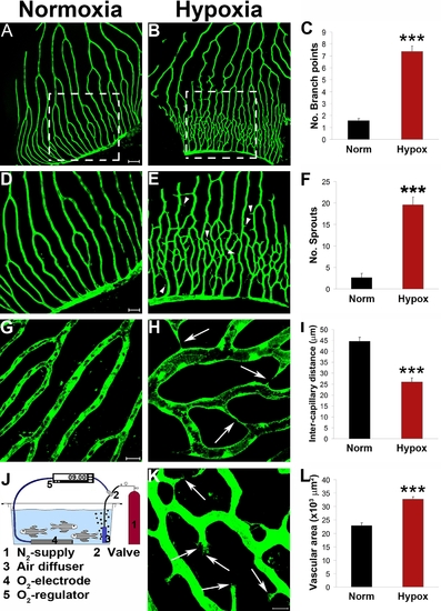
Hypoxia-induced retinal angiogenesis in adult fli-EGFP-Tg zebrafish. Adult fli-EGFP-Tg zebrafish were placed in a hypoxic aquaria and air saturation in the water was controlled at 10% (820 ppb) by an automated device (J). After 12-days exposure to this hypoxic environment, retinal angiogenesis in the capillary plexuses was detected (B, E, H, and K). Corresponding areas of the retinal vasculture exposed to normoxia were used as controls (A, D and G). Numbers of new vascular branches and sprouts, intercapillary distances, and total vascularization areas were accurately quantified (C, F, I, and L). Yellow arrowheads point to vascular sprouts. Yellow arrows point to endothelial tips. Data represents mean determinants of 11–16 randomized samples. ***p<0.001. Bar in panels A and B = 100 μm; in panels D and E = 50 μm; and in G and H = 20 μm. |