
Fig. 2
- ID
- ZDB-FIG-080529-67
- Publication
- Zhu et al., 2003 - Cloning, expression, and characterization of a membrane progestin receptor and evidence it is an intermediary in meiotic maturation of fish oocytes
- Other Figures
- All Figure Page
- Back to All Figure Page
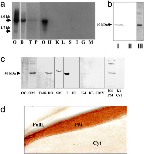
Tissue distribution and cellular localization of putative mPR. (a) Northern blot analysis showing mRNA expression in ovarian (O, gel loading: 1 μg) and other tissues (gel loading: 5 μg); B, brain; T, testis; P, pituitary; H, heart; K, kidney; L, liver; S, stomach; I, intestine; G, gill; M, muscle. (b) Western blot analysis of solubilized ovarian membrane proteins using monoclonal antibody PR10-1. I, plasma membrane; II, cytosol; III, partially purified membrane fraction used to generate monoclonal antibodies. (c) Cellular localization of receptor by Western blot analysis using stmPRαpAb1 antibody (gel loading: 10 μg). OC, oocyte cytosol; OM, oocyte membrane; Folli., follicle cell membrane; DO, denuded oocyte plasma membrane; SM, sperm membrane; I, recombinant protein induced by IPTG in E. coli; UI, noninduced E. coli protein; K4, membrane proteins from mPR-transfected MDA-MB-231 cells; K3, control cells transfected with vector containing reverse insert; CMV, control cells transfected with empty carrier vector. The following lanes were probed with PR10-1 antibody: K4 PM, plasma membrane from mPR transfected MDA-MB-231 cells; K4 Cyt, cytosol from mPR transfected cells. (d) Immunocytochemical localization of mPR receptor protein in a mature seatrout follicle containing a stage IV oocyte using stmPRαpAb1 antibody. Folli., follicle cells; PM, oocyte plasma membrane; Cyt, oocyte cytoplasm. (Magnification: 1 cm ≈ 20 μm.) |