
Fig. S5
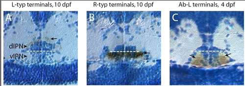
Localization of habenular axon arbors to the dIPN or vIPN. (a-c) Transverse plastic sections through the IPN of larvae in which habenular neurons were labeled by focal electroporation, followed by anti-GFP immunostaining (brown) to determine the location of axon arbors (indicated by arrows). (a) L-typical arbors are localized in the neuropil surrounding and covering the dIPN. (b, c) By contrast, R-typical terminals (b) and Ab-L terminals (c) are located in the vIPN. For (c), the parapineal ablation was performed in a Tg(flh:eGFP); Tg(foxD3:GFP) transgenic embryo in which GFP is weakly expressed in the habenular axons innervating the vIPN. The strongly labeled Ab-L terminals are seen as dark puncta (arrows in (c)) within the more lightly stained vIPN neuropil. Panels show transverse sections through 10 dpf (a, b) or 4 dpf (c) larval brains. Dotted white lines indicate the boundary between the dorsal and ventral parts of the IPN. |