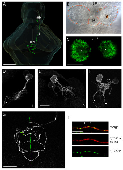
Habenular neurons have a stereotypical unipolar morphology and their axons terminate in spiral-shaped arbors that display multiple midline crossing. (a) Three-dimensional reconstruction showing a R habenular (rHb) projection neuron in an intact 4 dpf larval zebrafish brain. Arrow indicates direction of axonal projection within the FR, from the rHb to the IPN. (b) A single R habenular neuron labeled by focal electroporation and visualized by anti-GFP immunostaining (brown). The image shows the dorsal diencephalon of a dissected brain of a 4 dpf larva. Dotted lines show the borders of the habenulae and the position of the pineal stalk (PS). (c) Single-depth confocal section through the habenulae of a 4 dpf Tg(ET16:GFP) transgenic larva in which a subset of habenular neurons express GFP. In each nucleus the neuronal somata are arranged as ovoid shells surrounding a central neuropil domain (asterisks). It is in this domain that neurons elaborate their dendritic arbors. (d-f) Examples of the somata, dendritic arborizations and proximal axons of habenular neurons labeled by focal electroporation of membrane-tethered GFP. (d, e) Neurons with long processes that give rise to a dendritic tree and axon. (f) Two neurons with intertwined dendritic arbors close to the cell body. In (d-f) an arrowhead marks the proximal axon, and the laterality (left (L) or right (R)) of the habenular neuron(s) is indicated bottom right. (g) Confocal z-projection within the IPN showing a single axonal arbor elaborated by a R habenular projection neuron labeled by focal electroporation. The arbor crosses the ventral midline (dotted line) multiple times. Branches can also reverse direction such that they encircle the IPN in opposite senses (green arrows show examples). (h) High-magnification images of a section of a habenular axon arbor crossing the ventral midline (dotted line). The neuron was electroporated with a construct driving expression of cytosolic DsRed (red, middle panel) and a Syp-GFP fusion protein (green, lower panel). Syp-GFP puncta are present on both sides of the midline and co-localise with axonal variscocities. Scale bars: (a) 100 μm; (c) 50 μm; (e, h) 10 μm; (g) 20 μm.
|
