
Fig. 6
- ID
- ZDB-FIG-080506-34
- Publication
- Cole et al., 2001 - Apoptosis in the developing zebrafish embryo
- Other Figures
- All Figure Page
- Back to All Figure Page
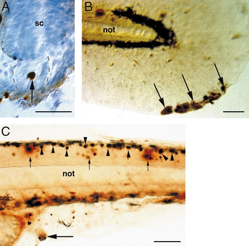
Apoptosis in other structures. (A) Transverse section of notochord at 20 hpf; dorsal is up. Arrow indicates large apoptotic cell within the notochord (outlined with dotted lines). (B) Whole-mount tail of 60-hpf embryo; anterior is left and dorsal is up. Arrows indicate large apoptotic cells at the distal margin of the median fin fold. (C) Lateral view of trunk of 48-hpf whole-mount embryo; anterior is left, dorsal is up. Muscle, spinal cord, and vent all contained apoptotic cells. Small arrows indicate apoptotic cells within the epaxial muscle. Arrowheads indicate apoptotic Rohon–Beard cells within the spinal cord. Large arrow indicates cluster of apoptotic cells at the prospective vent. Abbreviations: not, notochord; sc, spinal cord. Scale bars, 50 μm in (A–C). |
Reprinted from Developmental Biology, 240(1), Cole, L. and Ross, L., Apoptosis in the developing zebrafish embryo, 123-142, Copyright (2001) with permission from Elsevier. Full text @ Dev. Biol.