
- Title
-
Apoptosis in the developing zebrafish embryo
- Authors
- Cole, L. and Ross, L.
- Source
- Full text @ Dev. Biol.

General appearance and dynamics of apoptosis in whole-mount embryos. (A, B) Apoptotic cells are rapidly eliminated. At 20 hpf (A), the tailbud (region between arrows) demonstrates high levels of apoptosis. By 22 hpf (B), the tail region demonstrates little apoptosis. (C) Regions undergoing apoptosis typically contained both rounded (C, arrow) and shrunken, or pyknotic cells (C, arrowheads). Box in (A) demonstrates the region illustrated at higher power in (C). Rostral is left and dorsal is up. Scale bar in (A), 50 μm, applies to (A, B). Scale bar in (C), 5 μm. |
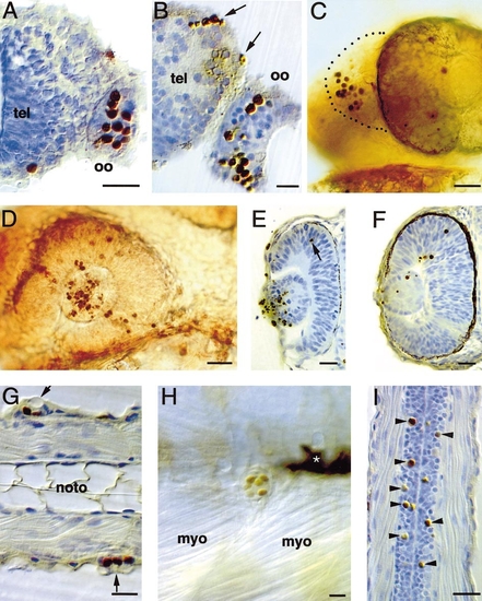
Apoptosis in sensory organs. (A–C) Olfactory organ. (A, B) Transverse sections of 36- (A) and 48-hpf (B) embryos; dorsal is up and lateral is right. (C) Whole-mount of 36-hpf embryo; rostral is left and dorsal is up. (A) The anterior surface of the olfactory organ contains numerous apoptotic cells at 36 hpf. (B) At 48 hpf, the morphology and location of apoptotic cells in intermediate layers suggest that they are dying neurons. Apoptosis is also seen at the margin of the telencephalon, in the prospective olfactory bulb region (arrows). (C) Lateral view of the head at 36 hpf demonstrates clusters of dying cells in the rostral olfactory organ (dotted lines). (D–F) Eye. (D) Whole-mount lateral view of eye at 24 hpf shows large numbers of apoptotic cells in the lens and scattered apoptotic cells in the retina at this age. Rostral is left and dorsal is up. (E) Transverse section at 24 hpf demonstrates that the majority of the apoptotic cells in the lens are at the outer margin, while the retina contains few apoptotic cells (arrow). Lateral is left and dorsal is up. (F) Transverse sectin at 36 hpf reveals numerous apoptotic cells in the retina at various levels and fewer apoptotic cells in the lens than seen at 24 hpf (compare to E). Lateral is left and dorsal is up. (G, H) Neuromasts of trunk lateral line organs. (G) Horizontal section of 36-hpf embryo, showing apoptotic cells within lateral line organs on outer surface of trunk (arrows). (H) Lateral view of single lateral line organ in 72-hpf whole mount; large apoptotic cells are clustered in the central region of the organ. Asterisk indicates pigment cells. Dorsal is up. (I) Apoptotic Rohon–Beard neurons (arrowheads) in a horizontal section of the dorsal spinal cord of a 48-hpf embryo. Rostral is up. Abbreviations: myo, myotome; noto, notochord; oo, olfactory organ; tel, telencephalon. Scale bars, 25 μm for all panels. |

Apoptosis in sensory organs and brain regions. (A–C) Transverse sections through inner ear at 36 hpf (A, B) and 60 hpf (C); dorsal is up and lateral is left. (A) The anterior macula and the statoacoustic ganglion contained apoptotic cells at 36 hpf. (B) The posteromedial macula also contained apoptotic cells at 36 hpf. (C) The semicircular canal and posterior macula contained apoptotic cells at 60 hpf. (D) Lateral view of 36 hpf whole mount; anterior is left and dorsal is up. Apoptosis in the trigeminal ganglion (arrow), anterior lateral line ganglion (arrowhead) and otocyst (asterisk). (E–H) Brain region apoptosis in transverse sections; dorsal is up in all panels. (E) At 48 hpf, apoptotic cells in the telencephalon were frequently located adjacent to the olfactory placode, in the region where ingrowing axons terminate. (F) Apoptotic cells at 48 hpf in the pineal gland (arrow). (G) Hypothalamus at 60 hpf. Throughout development, numerous apoptotic cells (arrows) were observed in the hypothalamus. (H) Tectum and tegmentum of the midbrain. At 60 hpf, the tectum contained numerous apoptotic cells, often clustered symmetrically (arrows) but also as single cells (arrowheads). Apoptotic cells in the tegmentum were less frequent (asterisk). Abbreviations: am, anterior macula; di, diencephalon; ol, otolith; oo, olfactory organ; pm, posteromedial macula; sag, statoacoustic ganglion; sc, semicircular canal; tec, tectum; teg, tegmentum; tel, telencephalon. Scale bars in (A), 25 mm for (A) and (B); in (C), 25 μm for (C), (E), and (F); in (D), 25 μm for (D), (G), and (H). |
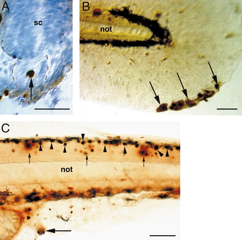
Apoptosis in other structures. (A) Transverse section of notochord at 20 hpf; dorsal is up. Arrow indicates large apoptotic cell within the notochord (outlined with dotted lines). (B) Whole-mount tail of 60-hpf embryo; anterior is left and dorsal is up. Arrows indicate large apoptotic cells at the distal margin of the median fin fold. (C) Lateral view of trunk of 48-hpf whole-mount embryo; anterior is left, dorsal is up. Muscle, spinal cord, and vent all contained apoptotic cells. Small arrows indicate apoptotic cells within the epaxial muscle. Arrowheads indicate apoptotic Rohon–Beard cells within the spinal cord. Large arrow indicates cluster of apoptotic cells at the prospective vent. Abbreviations: not, notochord; sc, spinal cord. Scale bars, 50 μm in (A–C). |
Reprinted from Developmental Biology, 240(1), Cole, L. and Ross, L., Apoptosis in the developing zebrafish embryo, 123-142, Copyright (2001) with permission from Elsevier. Full text @ Dev. Biol.