
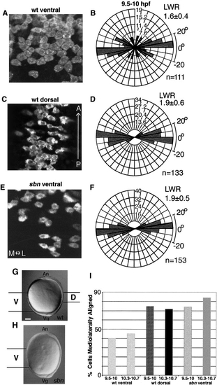
Fig. 6
Cells at low levels of Bmp activity are mediolaterally aligned and elongated. Confocal microscope images of DiI-labeled cells from (A) WT ventral, (C) WT dorsal, and (E) sbn ventral regions (9.5–10.3 hpf). (B, D) Rose diagrams of cell orientations from WT and (F) sbn embryos at 9.5–10 hpf. The mediolateral axis corresponds to the horizontal plane, while the anteroposterior axis aligns vertically. Cell number scale is indicated on each diagram. (G, H) An overview of the locations of cells analyzed in WT and sbn embryos. (I) Graph shows percentage of analyzed cells, which are mediolaterally aligned, i.e., orient the long axis ±20° with respect to the mediolateral axis. Hatched bars indicate late gastrula stages (9.5–10 hpf); solid bars are early segmentation stages (10.3–10.7 hpf). n, number of cells analyzed; LWR, length/width ratio. Scale bar, 100 μm. |
Reprinted from Developmental Biology, 243(1), Myers, D., Sepich, D., and Solnica-Krezel, L., Bmp activity gradient regulates convergent extension during zebrafish gastrulation, 81-98, Copyright (2002) with permission from Elsevier. Full text @ Dev. Biol.