
Fig. 4
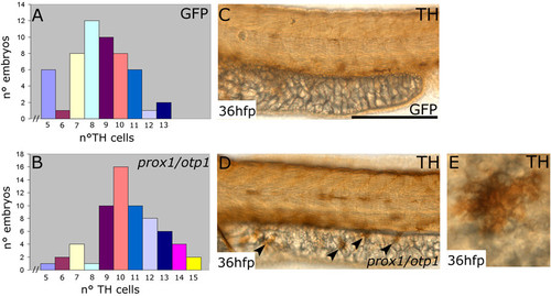
Synergistic prox1/otp1 overexpression induces the appearance of hypothalamic supernumerary TH-positive neurons and ectopic TH-positive cells on the yolk surface ectoderm. Lateral view, anterior is left and dorsal is up. (A,B) Distribution of TH positive cells in control and overexpressed prox1/otp1 embryos at 36 hpf. (A) The most numerous class in the group of GFP mRNA injected control embryos presented 8 CA hypothalamic neurons, and only 3 embryos presented more than 11 TH hypothalamic positive cells (n = 54). (B) The most numerous class in the group of the overexpressed prox1/otp1 embryos (n = 64) presented 10 CA neurons, and 20 embryos showed more than 11 TH hypothalamic positive cells. (C,D) Immunostaining with TH antibody shows ectopic TH positive cells on the yolk surface ectoderm of prox1/otp1 double injected embryos (arrowheads), while these cells are not present on the yolk of control embryos. (E) Ectopic TH positive cell on the yolk surface ectoderm. Scale bars indicate 50 μm. |