
Fig. 3
- ID
- ZDB-FIG-080328-12
- Publication
- Bit-Avragim et al., 2008 - Nuclear localization of the zebrafish tight junction protein nagie oko
- Other Figures
- All Figure Page
- Back to All Figure Page
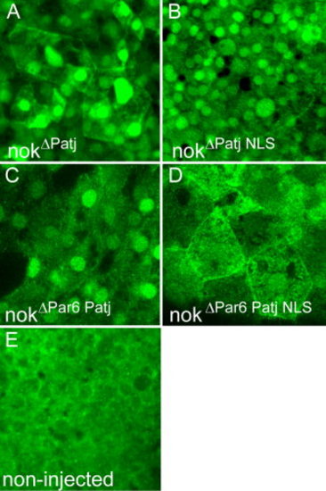
Redundant mechanisms involved in nok nuclear accumulation. Images are reconstructions of confocal Z-stack sections imaged on late gastrula stage whole mounts. HisMyc-tagged nok deletion proteins are detected with anti-Myc antibody (green). A: HisMyc-tagged nokΔPatj strongly localizes to nuclei of EVL and DL cells (recognizable by their size) and to outer cell membranes. B: HisMyc-tagged nokΔPatj NLS protein that lacks the L27N domain and the two nuclear import signals still localizes to the nucleus, which indicates that alternatives routes of nok nuclear import must exist. C: HisMyc-tagged nok nokΔPar6 Patj protein localizes to nuclei of EVL and DL cells indicating that direct association with Par6 is not necessary for nuclear accumulation of nok. D: In contrast, HisMyc-tagged nokΔPar6 Patj NLS protein fails to localize to nuclei of EVL and DL cells suggesting redundancy of the NLS motifs and association with Par6 in nok nuclear accumulation. E: Control non-injected embryos stained with the anti-Myc antibody. |