
Fig. 4
- ID
- ZDB-FIG-080326-113
- Publication
- Raschperger et al., 2008 - The coxsackie and adenovirus receptor (CAR) is required for renal epithelial differentiation within the zebrafish pronephros
- Other Figures
- All Figure Page
- Back to All Figure Page
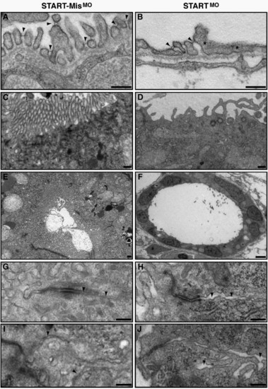
Ultrastructural analysis of CAR morphant pronephros reveals fused podocyte foot processes and abnormalities of the epithelium. Comparison of the ultrastructural morphology of control and CAR morphants at 96 hpf. (A and B) While the pronephric glomerulus of the control morphants (A) has well shaped and evenly spaced podocyte foot processes that are connected with slit diaphragms (arrowheads), the CAR morphants display a reduced number of slit diaphragms, as seen as fused podocyte foot processes (asterisks, B). The epithelial microvilli of the pronephric tubules are markedly reduced or missing in the CAR morphants (D and F), when compared to control morphants (C and E). In addition, CAR morphants display an increased distance between the neighboring plasma membranes (arrowheads) in pronephric tubular (H) and gut (J) epithelium, when compared to control morphants (G and I, respectively). Scale bar, 250 nm, except in panel F, 25 μm. |
| Fish: | |
|---|---|
| Knockdown Reagent: | |
| Observed In: | |
| Stage: | Day 4 |
Reprinted from Developmental Biology, 313(1), Raschperger, E., Neve, E.P., Wernerson, A., Hultenby, K., Pettersson, R.F., and Majumdar, A., The coxsackie and adenovirus receptor (CAR) is required for renal epithelial differentiation within the zebrafish pronephros, 455-464, Copyright (2008) with permission from Elsevier. Full text @ Dev. Biol.