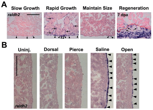
Developmental activation of the epicardium in response to growth or manipulation of the extracardiac environment. (A) Ventricles from SG, RG, or MS animals stained for raldh2 expression. raldh2 is weakly expressed in rare cells of SG and MS epicardia (arrowheads), but strongly expressed in epicardial cells of RG hearts, similar to the expression seen at the wound at 7 days post-amputation (right). Ventricular endocardial cells surrounding inner trabecular myofibers also induced raldh2 during rapid growth, similar to induction during regeneration (arrows in RG and Regeneration). (B) Assessment of raldh2 expression in ventricular epicardium 3 days after different manipulations in 6-month-old MS animals (lateral ventricular wall is shown). Surgically opening the pericardial sac (Open) or injecting saline into the pericardial sac (Saline) stimulated raldh2 expression (arrowheads), whereas injuries to the dorsal side of the animal (Dorsal), or piercing the pericardial sac with an empty needle (Pierce) did not. Scale bars: 100 μm.
|
