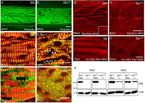
slou45 mutants have disorganised or missing expression of sarcomeric proteins. (A-J) Lateral views of muscle fibres in wild-type (Sib) (A,C,E,G,I) and slou45 (B,D,F,H,J) zebrafish embryos of ages shown bottom left and with reagents/antibodies shown bottom right. (A,B) F-Actin labelling with phalloidin showing a regular arrangement of fibrils in wild-type muscle fibres, whereas the slou45 mutant fibrillar organisation is disrupted. (C,D) Immunohistochemistry for α-Actinin (green) marks the Z-disc, here combined with phalloidin (red); the merge appears yellow. Z-discs are present in the slou45 mutant (white arrowheads, D) but are disordered: the distance between Z-discs is irregular (blue arrowheads) and Z-discs from neighbouring fibrils are not in register with each other (inset, D). Z-discs are flanked by Actin filaments in both siblings and mutants. (E,F) Titin labelling (green) using an antibody that marks a region of the molecule around the Z-disc, counterstained with phalloidin (red). (G-J) Immunohistochemistry for MHC (slow muscle Myosin, F59, red in G,H; pan-Myosin A4.1025, red in I,J). In the slou45 muscle cells, anti-MHC staining is severely reduced and lacks organisation. (K) Western blots using anti-MHC antibody F59 and γ-Tubulin (γ-Tub) as a loading control on lysates of slou45 and slotu44c mutant and sibling embryos at 30 hpf and 48 hpf. MHC protein levels are reduced in mutants of both alleles. Scale bars: 20 μm in A,B,G,H; 10 μm in C-F; 8 μm in I,J.
|
