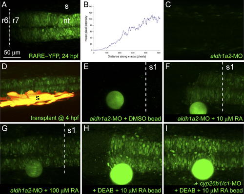
Responses of Embryos to Sources of RA. Long-range induction of a rare:yfp reporter. Confocal images of live embryos, dorsal view, anterior to the left. (A) rare:yfp expression in posterior hindbrain and spinal cord at 24 hpf with an anterior boundary at r6/7. (B) Quantification of fluorescence intensities in (A). (C) Lack of YFP expression in an aldh1a2 morphant embryo. (D) Rescue of YFP expression in a morphant by somitic mesoderm (yellow) transplanted during gastrulation. (E–I) Bead implantation. RA-coated beads were implanted anterior to the first somite at 18–19 hpf and imaged at 23–24 hpf. A DMSO-coated bead (E) failed to rescue YFP expression, whereas beads soaked in either 10 μM (F) or 100 μM (G) RA induced YFP at distances of up to 200 μm. (H and I) Inhibition of both cyp26b1 and cyp26c1 in DEAB-treated embryos leads to a symmetrical response from the rare:yfp reporter to a bead soaked in 10 μM RA (compare [H] with [I]). The dotted line indicates the r6/7 boundary, the dashed line, the anterior border of somite 1. nt, neural tube; s, somites.
|
