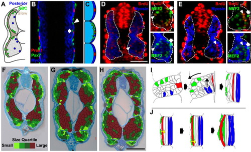
Distinct waves of myogenesis and a model for myotome development. (A) A summary of the fates of injected ABCs and posterior cells at 24 hours. Some injected ABCs (green) have formed differentiated fast muscle fibres, all are between superficial slow fibres and the earlier formed, fast fibres. All posterior cells (blue) have differentiated into medial fast fibres. (B) Dorsal view of a 24-hour embryo labeled for Pax7 (green), Prox1 (red) and myosin (blue). Slow fibre nuclei (red, Prox1) are at the surface of the myotome (blue, myosin). Most Pax7-positive nuclei (green) are outside of the myotome, but some are within the slow (arrowhead) or fast (arrow) domains of the myotome. (C) Schematic showing the location of Pax7-positive nuclei that are within the myotome. The pax7-positive nuclei within the myotome (MyHC-positive domain) from 30 different somites (somites 9-11 in a 24-hour embryo) are shown. (D) Transverse section through a mid-trunk somite (S9-12) of a 24-hour embryo treated with BrdU from the 5S stage (11 hours 40 minutes) to 24 hours. BrdU-positive nuclei (red, arrowheads) are found throughout the myotome (MyHC, blue), except for the superficial slow nuclei (arrows) (Barresi et al., 2001). Insets show muscle nuclei labeled with MEF2 (green). (E) Transverse section through a mid-trunk somite (S9-12) of a 24-hour embryo treated with BrdU from the 20S stage (19 hours) to 24 hours. BrdU-positive nuclei in the myotome are found solely within the lateral fast myotome (arrowheads). (F-H) Fibres with a smaller cross-sectional area are found laterally at 48 hours (F), 72 hours (G) and 96 hours (H); superficial slow fibres were excluded from this analysis. Relative cross-sectional area is shown according to size quartiles: light-green fibres, smallest 25%; dark-red fibres, largest 25%. (I,J) Models of somite cell movements and fates, viewed from dorsal (the midline is to the right). (I) Posterior cells (blue) become the medial fast fibres and ABCs (green) move laterally, becoming external cells expressing Pax7. (J) External cells then move medially into the myotome to become lateral fast fibres. During this time, adaxial cells (red) are displaced laterally and become superficial slow muscle fibres. Scale bars: 50 μm in D,H.
|
