Fig. 7
- ID
- ZDB-FIG-071112-15
- Publication
- Luo et al., 2007 - Inca: a novel p21-activated kinase-associated protein required for cranial neural crest development
- Other Figures
- All Figure Page
- Back to All Figure Page
|
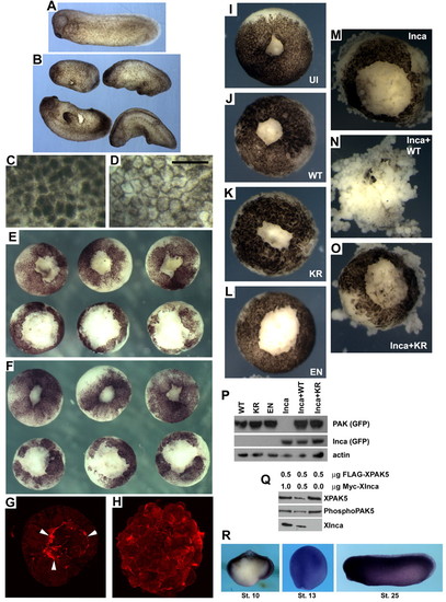
Inca misexpression disrupts gastrulation, pigmentation and wound healing in Xenopus. (A) Uninjected stage 25 control embryo. (B) Siblings injected with 250 pg IncaA mRNA have shorter body axes and numerous protuberances. (C,D) High-magnification views of ectoderm at stage 8 from uninjected (C) and IncaA mRNA-injected (250 pg; D) embryos showing relocation of cortical pigment to the cell periphery. Scale bar: 100 μm. (E,F) Vegetal explants after animal cap removal and culture for 5 (E) or 25 (F) minutes in 0.3xMMR. Upper row, uninjected; lower row injected with 250 pg IncaA mRNA, showing delayed healing. (G,H) Confocal z-stack images of animal caps from uninjected (G) and IncaA mRNA-injected (H) embryos after 5 minutes of healing in 0.3xMMR. White arrowheads in G indicate purse-string actin-filament structures missing in embryos overexpressing Inca. (I-O) Synergistic effect on wound healing of PAK5 and Inca. Embryos injected with mRNA encoding wild-type PAK5-GFP (J), kinase-dead PAK5-GFP (PAK5/KR; K), constitutively active PAK5-GFP (PAK5/EN; L), IncaA-GFP (M), a mixture of IncaA-GFP and PAK5-GFP (N) or a mixture of IncaA-GFP and PAK5/KR-GFP (O). All PAK5-GFP mRNAs injected at 1 ng, IncaA-GFP mRNA at 250 pg. Vegetal explants were allowed to heal for 25 minutes. Combining Inca+wild-type PAK5 greatly delays wound healing and leads to dissociation (N). PAK5/KR does not exhibit this synergistic effect (O). (I) Uninjected control embryo. Results from these experiments are summarized in Table 1. (P) PAK5-GFP mRNAs were equally translated, as judged by western blot using antibody to GFP. (Q) Co-expression with Inca does not increase PAK5 phosphorylation. Western blots of extracts made from HEK293 cells transfected with 0.5 µg FLAG-tagged XPAK5 plasmid and 0, 0.5 or 1.0 µg of Myc-tagged Xenopus Inca. The ratio of total XPAK5 signal (FLAG antibody) to the level of phosphorylation of regulatory serine residue 474 (phospho-PAK4) was not significantly different in any of the samples. (R) Xenopus PAK5 expression. Whole-mount in situ hybridization at stages 10, 13 and 25. Stage 10 embryos were bisected in the sagittal plane prior to hybridization. PAK5 expression is broad in ectoderm and mesoderm at early stages and essentially ubiquitous later in development. |

