
Fig. 6
- ID
- ZDB-FIG-071112-14
- Publication
- Luo et al., 2007 - Inca: a novel p21-activated kinase-associated protein required for cranial neural crest development
- Other Figures
- All Figure Page
- Back to All Figure Page
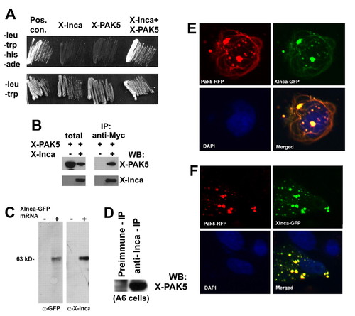
Inca interacts with PAK5. (A) Selective medium streaks of yeast cells expressing Xenopus Inca and PAK5, either alone or together, as indicated. Inca+PAK5 allows growth on medium lacking leucine, tryptophan, histidine and adenine. Positive controls include transfection with vectors containing T antigen and p53 (Clontech). Streaks on -leu/-trp (lower panel) shown as a control for transfection. (B) Extracts prepared from HEK293 cells transfected with expression plasmids encoding a PAK5-GFP fusion or a Myc epitope-tagged Inca separately or together, immunoprecipitated with anti-Myc, followed by western blot with antibody for GFP or Myc. Lanes labeled as total are from lysates prior to immunoprecipitation. PAK5-GFP precipitates with the anti-Myc antibody, but only when Myc-tagged Inca is present in the extract. (C) Anti-IncaA peptide antibody specificity. Fertilized eggs were injected with 1 ng of synthetic mRNA encoding XInca-GFP, and cultured to stage 20. Detergent (1% NP40)-solubilized protein was extracted with 1,1,2-trichlorotrifluoroethane (Freon) to remove yolk and the equivalent of two embryos analyzed by SDS-PAGE/western blot. Both anti-GFP and anti-Inca recognized a single band of the correct molecular weight (∼63 kDa). (D) Extract from untransfected Xenopus A6 cells immunoprecipitated with preimmune serum or anti-Inca, followed by western blot using antiserum raised against PAK5 (residues 122-224, a generous gift of N. Morin). Immunoprecipitation with anti-Inca enriches the PAK5 signal several-fold compared with preimmune serum, indicating Inca-PAK5 interaction. (E) Fluorescent images of a CHO cell cotransfected with plasmids encoding a PAK5-RFP fusion and Xenopus Inca-GFP fusion showing extensive overlap. (F) Fibrous structures positive for Inca and PAK5 are nocodazole-sensitive. CHO cells transiently transfected with PAK5-RFP and XInca-GFP were treated for 1 hour with 3 ng/ml nocodazole (Sigma), then fixed with methanol and photographed using an inverted fluorescence microscope. The fibers visible without nocodazole treatment (E) have disappeared. |