
Fig. 3
- ID
- ZDB-FIG-070928-17
- Publication
- Hadrys et al., 2006 - Conserved co-regulation and promoter sharing of hoxb3a and hoxb4a in zebrafish
- Other Figures
- All Figure Page
- Back to All Figure Page
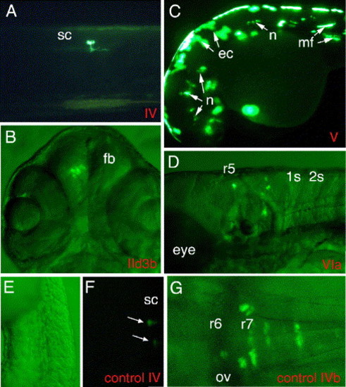
Reporter gene expression driven by hoxb3a and hoxb4a promoter sequences. (A) Construct IV including CNR1 sequences that are located directly upstream of shared exon 1 (hoxb3a and hoxb4a P2; Figs. 1A and 2) displayed weak basal promoter activity within the trunk in single mesodermal and neuronal cells; larva 72 hpf is shown. (B) EGFP reporter gene activity driven by hoxb3a P1 (construct IId3b) within the forebrain of a 72-hpf larvae. Combined fluorescent and bright field illumination. (C) An embryo of 48 hpf injected with the hoxb4a P1 reporter construct (V) is shown. This construct included a 1kb sequence immediately upstream of hoxb4a exon 2 (hoxb4a transcript I; Fig. 1 and Fig. 2) and displayed strong promoter activity. Mosaic EGFP expression was observed in neuronal, mesodermal, and epithelial cells throughout the whole embryo. (D) A sequence located within CNR11, directly upstream of hoxb3a exon 2 (Fig. 1 and Fig. 2), revealed promoter activity and drove EGFP expression in the posterior hindbrain derived from r5 through r7 and r8 and spinal cord. Combined fluorescent and bright field illumination is documented. (E–G) To confirm that the enhancers act through the endogenous hoxb3a/hoxb4a promoters and not through the E1b promoter located 52 of the EGFP coding sequence, we removed the Gal4VP16/UAS/E1b cassette from constructs IV (hoxb3a/hoxb4a P2) and VIb (hoxb3a P3-CNR10/CNR11 enhancer). Injection of these constructs resulted in much lower EGFP levels compared to the Gal4/UAS constructs, resulting in a lower rate of total EGFP expressing embryos and in a lower rate of neuronal expression (compare in Table 3). Panel E is bright field to panel F. (F) Embryo 24 hpf, injected with control construct IV that has EGFP label within the spinal cord. (G) Larva 48 hpf injected with control construct VIb. Abbreviations: ov, otic vesicle; r, rhombomere; sc, spinal cord. |
Reprinted from Developmental Biology, 297(1), Hadrys, T., Punnamoottil, B., Pieper, M., Kikuta, H., Pezeron, G., Becker, T.S., Prince, V., Baker, R., and Rinkwitz, S., Conserved co-regulation and promoter sharing of hoxb3a and hoxb4a in zebrafish, 26-43, Copyright (2006) with permission from Elsevier. Full text @ Dev. Biol.