
Fig. 2
- ID
- ZDB-FIG-070928-16
- Publication
- Hadrys et al., 2006 - Conserved co-regulation and promoter sharing of hoxb3a and hoxb4a in zebrafish
- Other Figures
- All Figure Page
- Back to All Figure Page
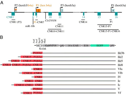
Illustration of PCR-amplified genomic fragments and expression vector design. (A) Locations of conserved non-coding sequences within the hoxb3a/hoxb4a cluster are indicated in turquoise, hoxb4a exons are in orange, and hoxb3a exons in light blue. The first exon is shared between hoxb3a and hoxb4a. Promoters (P) of hoxb3a and hoxb4a are indicated through arrows. Amplified sequences including different CNRs are illustrated below the schematic drawing. The retroviral insertion of zebrafish line CLGY838 and location of micro-RNA miR-10b are also indicated. (B) Fragments that were cloned in the Gal4VP16/UAS/E1b/EGFP expression vector and designation of the constructs. |
Reprinted from Developmental Biology, 297(1), Hadrys, T., Punnamoottil, B., Pieper, M., Kikuta, H., Pezeron, G., Becker, T.S., Prince, V., Baker, R., and Rinkwitz, S., Conserved co-regulation and promoter sharing of hoxb3a and hoxb4a in zebrafish, 26-43, Copyright (2006) with permission from Elsevier. Full text @ Dev. Biol.