
Fig. S1
- ID
- ZDB-FIG-070917-78
- Publication
- Dill et al., 2005 - tortuga refines Notch pathway gene expression in the zebrafish presomitic mesoderm at the post-transcriptional level
- Other Figures
- All Figure Page
- Back to All Figure Page
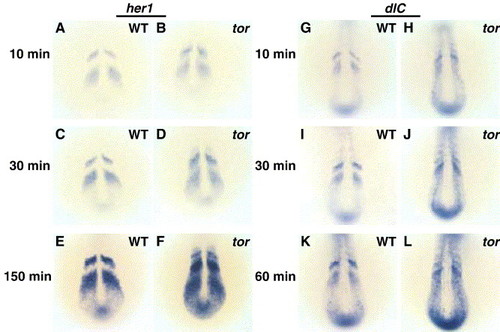
Ectopic expression of her1 and deltaC in tor mutant embryos is visible at times when wild type expression domains first become apparent. Embryos from the same clutch were fixed at 8 somites, hybridized with her1 (A–F) or deltaC (G–L) riboprobes, and processed in parallel coloration reactions. After 10 min, wild type expression domains are apparent (A, G). tor mutant embryos exhibit similar staining intensity in the wild type expression domains (stripes) with lower staining intensity in the ectopic interstripe domains (B, H). After longer incubations, both the wild type and ectopic expression domains show progressively darker staining intensity as the coloration reaction approaches saturation (B–F and H–L). Panels are dorsal views, anterior top. Genotype is indicated on each panel, and the duration of the coloration reaction is indicated to the left. Embryos were fixed before they could be separated by morphological criteria, and the ratio of presumptive wild type to tor mutant embryos was consistent with the expected 3:1 Mendelian ratio. |
Reprinted from Developmental Biology, 287(2), Dill, K.K., and Amacher, S.L., tortuga refines Notch pathway gene expression in the zebrafish presomitic mesoderm at the post-transcriptional level, 225-236, Copyright (2005) with permission from Elsevier. Full text @ Dev. Biol.