|
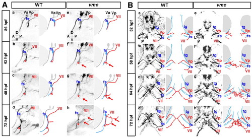
Time-lapse observations of the axonal outgrowth of the Vp and VII motoneurons in vmc embryos. (A) Time-lapse images of the axonal outgrowth of the VII motoneurons in wild-type (a-d) and vmc (e-h) zebrafish embryos. Lateral views; anterior, left. The axons of the VII motoneurons (red) and facial sensory (fs) neurons (blue) separated from each other at the points indicated by red asterisks (b-d,f-h). Arrows indicate the abnormal axons (f-h). (B) Time-lapse images of the axonal outgrowth of the Vp and VII motoneurons in wild-type (a-d) and vmc (e-h) embryos. Ventral views; anterior, top. The axons of the Vp and VII motoneurons (red) defasciculated into thin axons and grew abnormally (e-h). However, the axons of the trigeminal sensory (tg) and fs neurons (blue) grew normally (e-h).
|