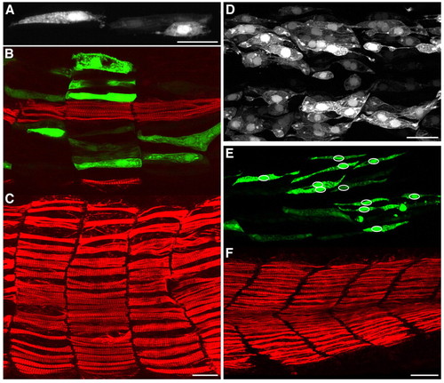
Morphant embryos show defective fast-myoblast fusion. (A) Embryo injected with dock5 splice 1 morpholino, showing examples in neighbouring somites of GFP-positive mononucleate cells that (left) span the somite and (right) have failed to elongate. (B,C) Embryo injected with dock5 splice 2 morpholino, showing an example of defects in somite shape. B is more medial and shows that the GFP-positive cells are medial to the superficial slow fibres shown in C, the slow fibres in B being the medially located muscle pioneers. (D) Embryo injected with crkl ATG morpholino, showing an example with many mononucleate GFP-labelled cells that have failed to elongate and still extend thin processes. (E,F) Embryo injected with dock5 splice 1 and crkl ATG morpholinos combined. Slow fibres are unaffected (F), although there are many mononucleate GFP-positive fast fibres (E). Circles in E indicate the positions of nuclei. All morpholinos were injected in solution containing mylz2:GFP plasmid and were fixed at 26-28 hpf. Green, GFP of mylz2:GFP plasmid; red, F59-antibody slow-fibre stain. Scale bars: 25 μm.
|
