
Fig. S1
- ID
- ZDB-FIG-070821-23
- Publication
- Murayama et al., 2005 - Otolith matrix proteins OMP-1 and Otolin-1 are necessary for normal otolith growth and their correct anchoring onto the sensory maculae
- Other Figures
- All Figure Page
- Back to All Figure Page
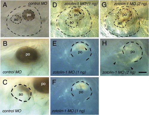
Spontaneous decalcification of the otoliths in fixed control and zotolin-1 MO-injected embryos. Embryos (four for each condition) were fixed at 7 dpf, kept in PBT at 4 °C, and observed 7 days later. All four control embryos still showed a complete posterior otolith and a portion of anterior otolith in both ears, as shown in (A–C). In contrast, 5/8 ears of the four embryos injected with 1 ng zotolin-1 MO had no visible mineralized otolith material left, and 3/8 had only a portion of posterior otolith, as shown in (D–F, arrowhead in D, E). Finally, 8/8 ears of embryos injected with 2 ng zotolin-1 MO had no mineralized otolith material left at all (G, H). Instead, sparse fiber-like material was spread through the otic cavity (arrowheads in H). Scale bar in (H) indicates 25 μm in (B, C, E, F, H). |
Reprinted from Mechanisms of Development, 122(6), Murayama, E., Herbomel, P., Kawakami, A., Takeda, H., and Nagasawa H., Otolith matrix proteins OMP-1 and Otolin-1 are necessary for normal otolith growth and their correct anchoring onto the sensory maculae, 791-803, Copyright (2005) with permission from Elsevier. Full text @ Mech. Dev.