
|
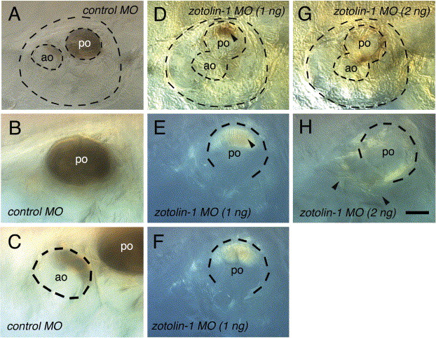
Fig. S1 Spontaneous decalcification of the otoliths in fixed control and zotolin-1 MO-injected embryos. Embryos (four for each condition) were fixed at 7 dpf, kept in PBT at 4 °C, and observed 7 days later. All four control embryos still showed a complete posterior otolith and a portion of anterior otolith in both ears, as shown in (A–C). In contrast, 5/8 ears of the four embryos injected with 1 ng zotolin-1 MO had no visible mineralized otolith material left, and 3/8 had only a portion of posterior otolith, as shown in (D–F, arrowhead in D, E). Finally, 8/8 ears of embryos injected with 2 ng zotolin-1 MO had no mineralized otolith material left at all (G, H). Instead, sparse fiber-like material was spread through the otic cavity (arrowheads in H). Scale bar in (H) indicates 25 μm in (B, C, E, F, H).
Reprinted from Mechanisms of Development, 122(6), Murayama, E., Herbomel, P., Kawakami, A., Takeda, H., and Nagasawa H., Otolith matrix proteins OMP-1 and Otolin-1 are necessary for normal otolith growth and their correct anchoring onto the sensory maculae, 791-803, Copyright (2005) with permission from Elsevier. Full text @ Mech. Dev.