
FIGURE
Fig. 2
- ID
- ZDB-FIG-070815-27
- Publication
- French et al., 2007 - Pbx homeodomain proteins pattern both the zebrafish retina and tectum
- Other Figures
- All Figure Page
- Back to All Figure Page
Fig. 2
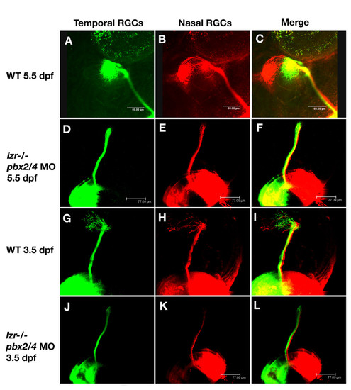
pbx2/4 null embryos exhibit RGC axonal outgrowth defects. In wild type embryos at 5.5 dpf, temporal RGC axons map to the anterior optic tectum (A) and (C), while nasal RGC axons map to the posterior region of the optic tectum (B) and (C). This is also seen at 3.5 dpf (G-I) although the connections on the tectum are more diffuse. In lzr-/- mutants injected with pbx2/4 morpholinos, both posterior and anterior RGC axons fail to map onto the optic tectum at both 3.5 dpf (J-L) and 5.5 dpf (D-F). |
Expression Data
Expression Detail
Antibody Labeling
Phenotype Data
| Fish: | |
|---|---|
| Knockdown Reagents: | |
| Observed In: | |
| Stage Range: | Protruding-mouth to Day 5 |
Phenotype Detail
Acknowledgments
This image is the copyrighted work of the attributed author or publisher, and
ZFIN has permission only to display this image to its users.
Additional permissions should be obtained from the applicable author or publisher of the image.
Full text @ BMC Dev. Biol.