
Fig. 5
- ID
- ZDB-FIG-070608-5
- Publication
- Hall et al., 2007 - The zebrafish candyfloss mutant implicates extracellular matrix adhesion failure in laminin {alpha}2-deficient congenital muscular dystrophy
- Other Figures
- All Figure Page
- Back to All Figure Page
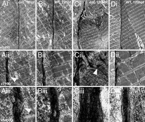
Transmission electron micrographs of the vertical myosepta in caf and wild-type embryos at 72 and 120 hpf. (A) caf, 72 hpf. The extracellular matrix phenotype is subtle and under low magnification (i; ×2,400) the myosepta appear normal. However, under higher magnification (ii; ×7,100 and iii; ×54,000), tearing of the myosepta can be seen that is not apparent in wild-type siblings (arrowheads). (B) Wild-type, 72 hpf. (C) caf, 120 hpf. Even under low magnification, the myosepta are grossly distorted and fibrotic. Under medium magnification, portions of extracellular matrix can be seen to infiltrate the myotome (arrowhead), pulled with the detaching fibers. Under high magnification, the myosepta are greatly increased in diameter and show condensed collagen fibers, indicative of a fibrotic response. (D) Wild-type, 120 hpf. |
| Fish: | |
|---|---|
| Observed In: | |
| Stage Range: | Protruding-mouth to Day 5 |