
Fig. 3
- ID
- ZDB-FIG-070418-41
- Publication
- Choi et al., 2007 - FoxH1 negatively modulates flk1 gene expression and vascular formation in zebrafish
- Other Figures
- All Figure Page
- Back to All Figure Page
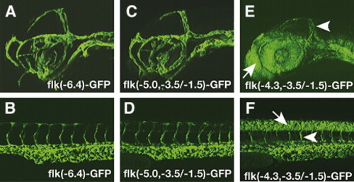
Transgenic analysis of the flk1 regulatory region identifies a 1.5-kb minimal endothelial specific enhancer. (A–B) GFP expression patterns in the brain (A) and trunk (B) of 2-day-old TG(flk1:GFP)la116 embryos. (C–D) GFP expression patterns of 2-day-old embryos carrying germ line integrated flk(-5.0, -3.5/-1.5)-GFP transgene resembles the patterns observed in TG(flk1:GFP)la116. (E–F) Embryos carrying germ line integrated flk(-4.3, -3.5/-1.5)-GFP have GFP expression in endothelial cells (arrowhead) as well as neural tissues. Arrows point to GFP positive cells in forebrain in panel E and to GFP positive cells in neural tube in panel F. |
Reprinted from Developmental Biology, 304(2), Choi, J., Dong, L., Ahn, J., Dao, D., Hammerschmidt, M., and Chen, J.N., FoxH1 negatively modulates flk1 gene expression and vascular formation in zebrafish, 735-744, Copyright (2007) with permission from Elsevier. Full text @ Dev. Biol.