
Fig. 2
- ID
- ZDB-FIG-070418-40
- Publication
- Choi et al., 2007 - FoxH1 negatively modulates flk1 gene expression and vascular formation in zebrafish
- Other Figures
- All Figure Page
- Back to All Figure Page
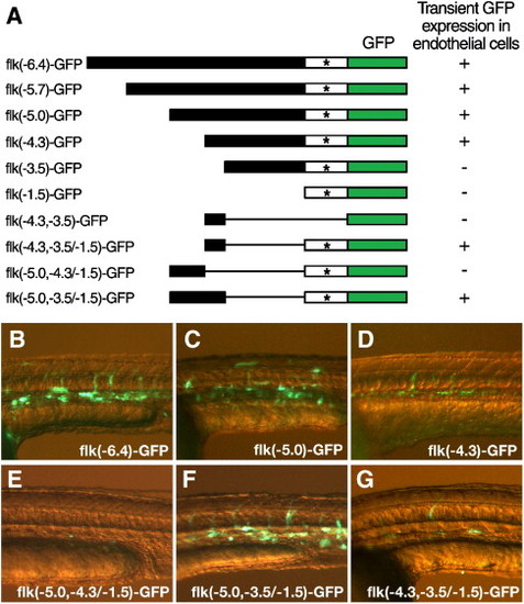
Deletion analysis of the flk1 regulatory region identifies a highly conserved element that is necessary for endothelial expression. (A) Schematic diagram of the deletion constructs of flk1-GFP reporter. Linearized DNA of each construct was injected in wild type zebrafish embryos at the 1-cell stage. GFP expression in injected embryos was analyzed after 1 day of development. The transient endothelial expression directed by each construct is summarized by a plus (endothelial expression) or a minus (no detectable endothelial expression) to the right of the line representing each construct. * marks the transcription initiation site of flk1. (B–G) Transient GFP expression in endothelial cells of 1-day-old embryos injected with flk(- 6.4)-GFP (B), flk(- 5.0)-GFP (C), flk(- 4.3)-GFP (D), flk(- 5.0, - 4.3/- 1.5)-GFP (E), flk(- 5.0, - 3.5/- 1.5)-GFP (F) or flk(- 4.3, - 3.5/- 1.5)-GFP (G). |
Reprinted from Developmental Biology, 304(2), Choi, J., Dong, L., Ahn, J., Dao, D., Hammerschmidt, M., and Chen, J.N., FoxH1 negatively modulates flk1 gene expression and vascular formation in zebrafish, 735-744, Copyright (2007) with permission from Elsevier. Full text @ Dev. Biol.