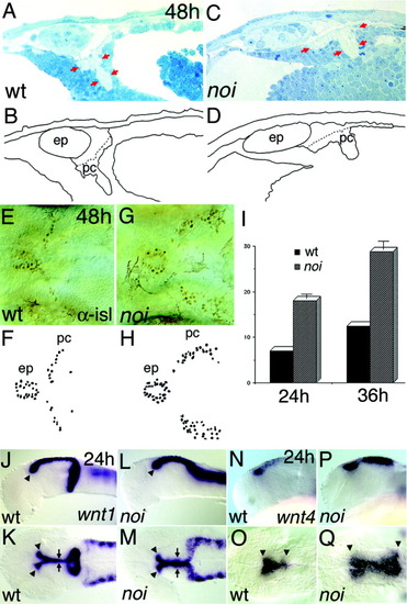
The territory of the dorsal forebrain and midbrain is altered in noi mutant embryos. Histologic, immunochemical, and in situ hybridisation (ISH) analysis was used to study the dorsal forebrain and midbrain in noi mutant embryos. The embryos were oriented with anterior to the left. A,B: A parasagittal section through the dorsal part of the forebrain-midbrain region of a 48 hours postfertilization (hpf; 48h) wild-type (wt) zebrafish embryo shows the location of the epiphysis (ep) and the posterior commissure (pc). C,D: In the mutant, the ep and the pc seem enlarged in the anteroposterior direction marked with red arrows in C. E-H: A dorsal view of the nucleus of the posterior commissure interneurons stained with an α-islet (αisl) antibody indicates that the number of neurons is increased and the territory is expanded posteriorly. I: Already at 24 hpf and at 36 hpf, a significantly increased number of neurons is observed between wild-type siblings and in noi mutant embryos. J-M: A lateral and dorsal view of an ISH of wnt1 shows a fusion of the midbrain and hindbrain pattern and the missing expression in the ventral part of the midbrain-hindbrain boundary. N-Q: The expression wnt4 shows an expansion of these dorsal diencephalic markers into the midbrain territory (arrowheads).
|
