
Fig. 5
- ID
- ZDB-FIG-061227-28
- Publication
- Lien et al., 2006 - Gene Expression Analysis of Zebrafish Heart Regeneration
- Other Figures
- All Figure Page
- Back to All Figure Page
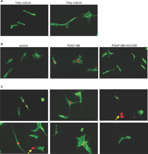
PDGF-B Induces DNA Synthesis in Adult Zebrafish Cardiomyocytes. (A) Adult zebrafish cardiomyocytes were isolated and cultured for 1 or 7 d. The cardiomyocyte population is heterogeneous in size and shape consisting mostly of rod-shape cells. To confirm that the cells are cardiomyocytes, the cells were stained with anti-tropomyosin antibody (green) and anti-MEF2 antibody (unpublished data). After 7 d in culture in 10% FBS, the cardiomyocytes underwent dedifferentiation; the cells lose their rod shape and striation becomes disorganized. (B) DNA synthesis as determined by BrdU incorporation in adult zebrafish cardiomyocytes treated with DMSO (control), PDGF-BB, and PDGF-BB plus the PDGF receptor inhibitor, AG1295. BrdU labeling is shown in red and tropomyosin staining is shown in green. PDGF-BB induced a 2.68-fold increase in DNA synthesis compared to control in adult zebrafish cardiomyocytes (p < 0.05). The effect of PDGF-BB inducing DNA synthesis can be blocked by AG1295. (C) PDGF-BB induced a variety of cardiomyocytes with different size and shape to undergo DNA synthesis. Noncardiomyocytes are marked by yellow arrows. |