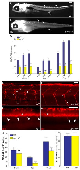
sox10 mutants display a DRG neuron phenotype. Immunofluorescent detection of Hu at 5 dpf (A,B), DM-GRASP (zn-5 antibody) at 8 dpf (D,E) and Isl-1 at 60 hpf (F,G) in wild-type (A,D,F) and sox10 (B,E,G) embryos demonstrates the reduction in DRG sensory neurons (arrowheads) in the mutant. Quantitation (mean+s.d.) of Hu (C) and Isl-1 (H) phenotypes shows increasing severity posteriorly. Significant reduction in sensory neurons is noted at all stages (** P<0.01, ***P<0.0001; Student's t-test). n=12 embryos for each of wild-type and sox10 mutants in (C) and n=15 (wild type) and n=13 (sox10) in (H). There is an absence of enteric neurons in sox10 mutants (arrow, B). However, DM-GRASP expression in secondary motorneuron processes (*,D) is unchanged in sox10 mutants (E). Likewise, Islet1 also labels ventral spinal cord motorneurons (*,F,G). Quantification (I) of these cells at 5 dpf per somite segment equivalent shows no significant difference between wild-type and sox10 embryos (P=0.609; Student's t-test). Three consecutive segment-lengths in the tail were counted in seven embryos of each genotype. Scale bars: 200 μm in A,B; 50 μm in D-G.
|
