
Fig. 4
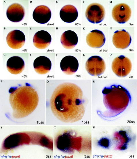
Early expression profiles of zebrafish sfrp1a. Dorsal with animal pole to the top (C, F, I, L and O), dorsal with anterior to the left (Q, T and U), lateral with anterior to the top (B, E, H, K, N, P, R and S) or animal pole with anterior to the top (A, D, G, J, and M) views of zebrafish embryos hybridized with sfrp1a antisense probe. S, T, and U are double in situ showing sfrp1a in blue and pax6 (S and T) or pax2 (U) in red. White asterisk indicates the pre- and somatic mesoderm. Abbreviations: a, anterior neural plate; h, presumptive hindbrain; m, presumptive midbrain; ov, optic vesicle; t, presumptive telencephalon; tb, tail bud. |
| Genes: | |
|---|---|
| Fish: | |
| Anatomical Terms: | |
| Stage Range: | 30%-epiboly to 20-25 somites |
Reprinted from Gene expression patterns : GEP, 6(8), Tendeng, C., and Houart, C., Cloning and embryonic expression of five distinct sfrp genes in the zebrafish Danio rerio, 761-771, Copyright (2006) with permission from Elsevier. Full text @ Gene Expr. Patterns