
Fig. 5
- ID
- ZDB-FIG-060901-19
- Publication
- Yang et al., 2006 - Small molecule-induced ablation and subsequent regeneration of larval zebrafish melanocytes
- Other Figures
- All Figure Page
- Back to All Figure Page
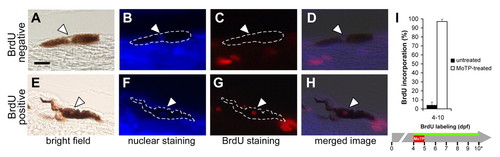
Larval melanocyte regeneration following MoTP treatment is achieved by cell division. (A-H) Cell division events in melanocyte lineages during larval melanocyte regeneration were tracked by BrdU incorporation experiments. Larvae were continuously incubated in BrdU (5 mM) during and after MoTP treatment, then fixed, paraffin embedded and 5 μm sagittal sections processed for BrdU immunohistochemistry after melanocyte regeneration was mostly completed at 10 dpf (5 days post-MoTP treatment). BrdU incorporation states of pigmented melanocytes were assessed by first identifying melanocyte nuclei (thinning in melanin, white arrowheads in A and E) accompanied by bisbenzimide staining (white arrowheads in B and F). These were then examined for red fluorescence indicative of BrdU incorporation (white arrowheads in C and G). D is the overlay image of A, B and C, indicating a melanocyte that did not incorporate BrdU during the BrdU labeling, whereas in H, the overlay image of D, E and F shows a different melanocyte that did incorporate BrdU during the BrdU labeling. (I) Quantitative analyses of BrdU incorporation in larvae exposed to MoTP from 4 to 5 dpf. Black and white bars indicate BrdU incorporation in untreated and MoTP-treated larvae, respectively. Horizontal green line indicates periods of BrdU labeling. Asterisk in I indicates the developmental stages at which larvae were sacrificed for BrdU incorporation analysis. Scale bar in A: 10 μm for A-H. |