
Fig. 8
- ID
- ZDB-FIG-060824-8
- Publication
- Gulati-Leekha et al., 2006 - A reporter-assisted mutagenesis screen using α1-tubulin-GFP transgenic zebrafish uncovers missteps during neuronal development and axonogenesis
- Other Figures
- All Figure Page
- Back to All Figure Page
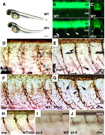
Motoxonal branching and terminal innervation is impaired in the dhv spinal cord. Mutants become discernible at 2 dpf displaying progressive paralysis and other dysmorphologies (bright-field images showing lateral view of embryos (A) accompanied by fluorescent ventral projections (arrows; B) from the spinal cord (sc, lateral view, anterior towards left) into the ventral myotome (vm). GFP immunostaining of transverse spinal cord sections further highlights the GFP-positive spinal nerves (arrowheads, C). znp-1 immunostaining of caudal (D, E) and rostral (F, G) myotomal segments at 57 hpf shows lack of secondary and tertiary (2′,3′; D) motoaxonal branches in dhv. Note the stunted lateral projections (arrows; panels E and G) and swollen varicosities (arrowheads; panels E and G). Intense staining of spinal cord precludes examination of dorsal myotome. Motor axonal pathfinding is indistinguishable between mutant and wild-type spinal cords at 32 hpf (representative znp-1 immunostaining; H). Mutation in dhruva forestalls down-regulation of DM-GRASP expression, which marks secondary motor neurons (smn) and corresponding fasciculated axonal segments (zn-5 immunostaining at 57 hpf, representative mid-trunk spinal cord segments; panels I and J). Scale bars: 250 μm in panel A, 50 μm in panel B, 25 μm in panels C, D–G, and H–J. |
| Gene: | |
|---|---|
| Fish: | |
| Anatomical Term: | |
| Stage: | Long-pec |
Reprinted from Developmental Biology, 296(1), Gulati-Leekha, A., and Goldman, D., A reporter-assisted mutagenesis screen using α1-tubulin-GFP transgenic zebrafish uncovers missteps during neuronal development and axonogenesis, 29-47, Copyright (2006) with permission from Elsevier. Full text @ Dev. Biol.