
Fig. 4
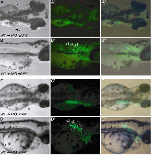
Mosaic analysis in raldh2 and prdm1 morphants. (A) Dorsal view of a three-day-old raldh2 morphant embryo revealing rescued pectoral fin outgrowth on the right side (arrow). (A') Dark-field image of the same embryo, showing transplanted GFP-positive cells labeled in green. (A'') Merged bright-field and dark-field images showing green wild-type cells localizing to the anterior somite region. (B-B'') Lateral views of the same MOraldh2 mosaic embryo as in A. Dotted lines in B' indicate somite boundaries. Note strong GFP-expression in somites 1 to 3. (C-D'') Dorsal and lateral views of an MOprdm1 embryo, where transplanted wild-type cells contribute to anterior somites but do not rescue fin outgrowth. (C'',D'') Merged bright and dark field pictures showing GFP-positive wild-type cells incorporated into the left fin. Dotted lines in D' indicate somite boundaries. Asterisks mark the missing pectoral fin. s, somite. |