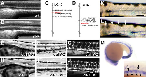
Characterization of somite patterning defects and molecular analysis of y56/y70 and y55/y66 mutants. A,B: Transmitted light micrographs of the trunks of 72 hours postfertilization (hpf) wild-type (A) and y56 mutant (B) animals, showing poor morphogenesis of somite boundaries in mutants. C,D: Genetic mapping of y56 and y70 (C) and y55 and y66 (D) mutants. C: y56 and y70 mutants are tightly linked to tbx24 on LG12. The numbers in parentheses show linkage (number of recombinants/total meioses) to CA repeat markers for y56 (first numbers in parentheses) and y70 (last numbers in parentheses). D: The y55 and y66 mutants are tightly linked to deltaC on LG15. The numbers in parentheses show linkage (number of recombinants/total meioses) to CA repeat markers for y55 (first numbers in parentheses) and y66 (last numbers in parentheses). NP indicates marker was not polymorphic for particular mutant and marker combination. E-L: Transmitted light (E,F,H,I) and TG(fli1:egfp)y1 vascular fluorescence (G,J) micrographs of 48 hpf trunks (E,G,H,J) and tails (F,I) of control (E-G) and deltaC (H-J) morpholino injected animals. H,I: Animals injected with deltaC morpholinos form somite boundaries 1-4 (H) but not more posterior boundaries (I) properly and show defects in vessel patterning indistinguishable from those seen in y55 and y56 mutants (J). K,L: Higher magnification transmitted light images of the anterior trunk of 3 days postfertilization wild-type (K) and y56 mutant (L) animals, with well-formed anterior somites in y56 (arrows). Whole-mount in situ hybridization of a 19-20 hpf wild-type animal using a probe for delC. Magnified inset shows expression in dorsal aorta (arrows) and somites (arrowheads). Anterior is to the left and dorsal is up in all micrographs. Scale bars = 100 μm.
|
