
Fig. 2
- ID
- ZDB-FIG-060424-1
- Publication
- Inoue et al., 2006 - Cloning and characterization of mr-s, a novel SAM domain protein, predominantly expressed in retinal photoreceptor cells
- Other Figures
- All Figure Page
- Back to All Figure Page
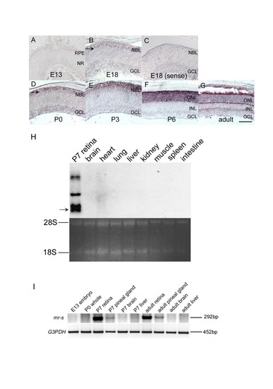
mr-s expression in developing mouse retina and pineal gland. (A-G) mr-s expression during development of the mouse retina. The in situ hybridization signal of mr-s was not detected at E13 (A). The signal (arrow) was first detected in the outer aspect of NBL at E18 (B). A strong mr-s signal was detected in outer layer of the retina at P3-P6, and then the signal decreased in the adult retina (E-G). Control with the sense probe in E18 retina is shown (C). Scale bar, 100 μm. (H) Northern blot analysis of mr-s expression in adult mouse organs. The arrow corresponds to 2.2kb mr-s transcript. (I) RT-PCR analysis of total RNAs extracted from E13 whole embryo, P0 whole body (except for the eye), P7 retina, P7 pineal gland, P7 brain, P7 liver, adult retina, adult pineal gland, adult brain and adult liver, respectively. RPE, retinal pigment epithelium; NR, neural retina; NBL, neuroblastic layer; GCL, ganglion cell layer; ONL, outer nuclear layer; INL, inner nuclear layer. |