
- Title
-
Cloning and characterization of mr-s, a novel SAM domain protein, predominantly expressed in retinal photoreceptor cells
- Authors
- Inoue, T., Terada, K., Furukawa, A., Koike, C., Tamaki, Y., Araie, M., and Furukawa, T.
- Source
- Full text @ BMC Dev. Biol.
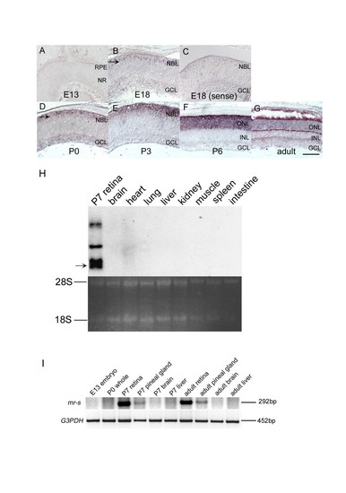
mr-s expression in developing mouse retina and pineal gland. (A-G) mr-s expression during development of the mouse retina. The in situ hybridization signal of mr-s was not detected at E13 (A). The signal (arrow) was first detected in the outer aspect of NBL at E18 (B). A strong mr-s signal was detected in outer layer of the retina at P3-P6, and then the signal decreased in the adult retina (E-G). Control with the sense probe in E18 retina is shown (C). Scale bar, 100 μm. (H) Northern blot analysis of mr-s expression in adult mouse organs. The arrow corresponds to 2.2kb mr-s transcript. (I) RT-PCR analysis of total RNAs extracted from E13 whole embryo, P0 whole body (except for the eye), P7 retina, P7 pineal gland, P7 brain, P7 liver, adult retina, adult pineal gland, adult brain and adult liver, respectively. RPE, retinal pigment epithelium; NR, neural retina; NBL, neuroblastic layer; GCL, ganglion cell layer; ONL, outer nuclear layer; INL, inner nuclear layer. |
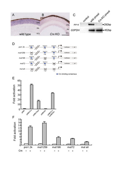
The transcription of mr-s is regulated by Crx. (A, B) In situ hybridization using a probe for mouse mr-s was performed on the wild-type (A) and Crx KO retinas (B) at P5. mr-s expression was drastically reduced in the Crx KO retina (B). Scale bar, 100 μm. (C) RT-PCR analysis of total RNAs extracted from the pineal glands of P5 wild-type and Crx KO mouse. The upper and lower lanes show PCR products amplified by the primer pairs specific for mr-s and G3PDH cDNAs, respectively. Water was used for control RT-PCR reaction. (D-F) Crx transactivates mr-s transcription. Reporter plasmids for the luciferase assay are shown. Blue boxes represent Crx binding sites (D). Relative activity of the luciferase is indicated when Pro1.2k was co-transfected with Crx, Otx2, Nrl, and Crx+Nrl, respectively (E). Fold activation is indicated when Pro1.2k, mut1259, mut198, mut72 and mut all were co-transfected with the Crx expression vector (Crx+) or the empty vector (Crx-) into HEK293T cells (F). Error bars represent standard error of mean. |

Subcellular localization of mr-s in mammalian cells. (A, B) HA-tagged full-length mr-s was transfected into HEK293T (B) and detected by anti-HA antibody (A). Scale bar, 20 μm. (C-E) HEK293T cells immunostained with anti-HA antibody (C), DAPI (D), and merged image (E). Scale bar, 50 μm. |