|
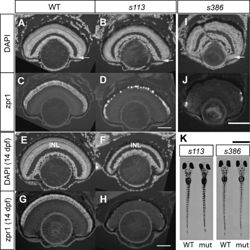
Examples of Mutants with Photoreceptor Degeneration. WT and mutant retinas (A-H, mtis113; I and J, ssds386) were sectioned and stained with DAPI (A, B, E, F, and I) and zpr1 monoclonal antibody (double-cone photoreceptor marker) (C, D, G, H, and J). At 7 dpf, photoreceptors in the central part of the retina have degenerated in both mti (A-D) and ssd (I-J). In the mti retina at 14 dpf, degeneration has spread to the inner nuclear layer (INL). Arrows show the ciliary marginal zone, from which new cells are continually added to the growing retina. Scale bar is 100 μm. (K) Mutants with photoreceptor degeneration may (mtis113) or may not (ssds386) be dark in VBA assay. Scale bar is 1 mm.
|

