
Fig. 6
- ID
- ZDB-FIG-051114-8
- Publication
- Julich et al., 2005 - beamter/deltaC and the role of Notch ligands in the zebrafish somite segmentation, hindbrain neurogenesis and hypochord differentiation
- Other Figures
- All Figure Page
- Back to All Figure Page
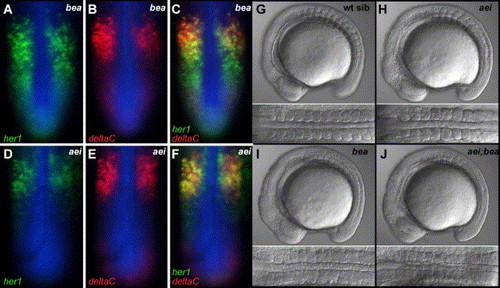
The oscillating expression patterns of her1 and deltaC are differentially affected in bea/deltaC and aei/deltaD mutants. (A–C) Show a bea/deltaCtit446 embryo stained for her1 mRNA (A, green), deltaC mRNA (B, red) and β-catenin protein (blue). (C) Overlay of A and B. The “salt and pepper” pattern of her1 extends more posteriorly than that of deltaC. (D–F) Show an aei/deltaDtg249 embryo stained for her1 mRNA (D, green), deltaC mRNA (E, red) and β-catenin protein (blue). (F) Overlay of D and E. The “salt and pepper” patterns of her1 and deltaC are both restricted to the anterior PSM. (G–J) Lateral and dorsal views of wild-type (G), aei/deltaDtg249 homozygotes (H), bea/deltaCtit446 homozygotes (I) and aei/deltaDtg249; bea/deltaCtit446 double homozygotes (J). At the 11 somite stage, there is no clear difference in the somite phenotype when comparing the bea/deltaC homozygotes to aei/deltaD; bea/deltaC double homozygotes. |
| Genes: | |
|---|---|
| Fish: | |
| Anatomical Term: | |
| Stage: | 10-13 somites |
Reprinted from Developmental Biology, 286(2), Julich, D., Hwee, Lim C., Round, J., Nicolaije, C., Schroeder, J., Davies, A., Geisler, R., Lewis, J., Jiang, Y.J., Holley, S.A., Tübingen 2000 Screen Consortium., beamter/deltaC and the role of Notch ligands in the zebrafish somite segmentation, hindbrain neurogenesis and hypochord differentiation, 391-404, Copyright (2005) with permission from Elsevier. Full text @ Dev. Biol.