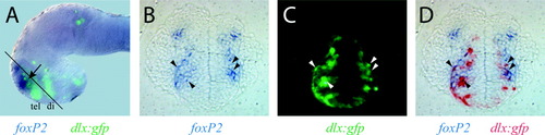
Co-expression of foxP2 and dlx5/6 in the subpallial telencephalon. dlx6a(156i, 156ii):gfp embryos at 32 hpf were double-stained for in situ expression of foxP2 (purple), and with an antibody directed against GFP and detected fluorescently using Alexa-488. A: Whole-mount embryo, lateral view of the head region with dorsal at the top (eyes removed for better visualization). foxP2 expression (purple) overlaps with the telencephalic band of dlx:gfp expression (green, tel) in a narrow area (arrow). Diencephalic expression of dlx:gfp is marked di. Approximate plane of the sections shown in B-D is indicated by the black line transecting the head. B-D: A single 7-μm transverse plastic section in the ventral telencephalon at the level of the rostral olfactory placode, dorsal is at top. Two arrowheads in each hemifield indicate representative cells that show co-expression. B: foxP2 in situ expression. C: GFP expression. D: Overlay of foxP2 and GFP expression, with GFP pattern shown using inverse contrast (in red).
|
