
Fig. 6
- ID
- ZDB-FIG-050916-6
- Publication
- Beis et al., 2005 - Genetic and cellular analyses of zebrafish atrioventricular cushion and valve development
- Other Figures
- All Figure Page
- Back to All Figure Page
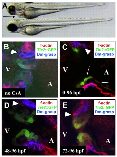
Calcineurin signaling is required for EMT and EC morphogenesis. (A) Bright-field image of an untreated embryo (bottom) and an embryo raised in 10 µg/ml CsA (top). (B-E) Confocal images of the AV canal of Tg(Tie2:EGFP)s849 (green) embryos at 96 hpf immunostained for Dm-grasp (blue) and stained with rhodamine phalloidine (red); (B) untreated embryo; (C-E) embryos treated with CsA from the one cell stage (C), 48 hpf (D) and 72 hpf (E). (A) Embryos treated with CsA from the one cell stage appeared morphologically wild type at 72 hpf, except for pericardial edema (arrow in A) owing to outflow tract stenosis (see Movie 1 in the supplementary material). (C) In embryos treated with CsA from the one-cell stage, the myocardium appeared thinner throughout the heart (compare with B, D, E and Fig. 2). AV endocardial cells upregulated Tg(Tie2:EGFP)s849 and initiated a cell shape change (thin arrow indicates AV endocardial cell, thick arrow indicates squamous atrial endocardial cell), but failed to express Dm-Grasp; no EMT occurred and ECs failed to form. (D) In embryos treated with CsA from 48 hpf, ECs appeared disorganized. (E) No effect on EMT or cushion morphogenesis was observed at 96 hpf when embryos were treated with CsA from 72 hpf onwards (compare E with B). Arrowheads indicate the superior region of the AV canal in B-E.
|
| Gene: | |
|---|---|
| Fish: | |
| Condition: | |
| Anatomical Term: | |
| Stage: | Day 4 |