
Fig. 2
- ID
- ZDB-FIG-050906-7
- Publication
- Chen et al., 2005 - A unique role for 6-O sulfation modification in zebrafish vascular development
- Other Figures
- All Figure Page
- Back to All Figure Page
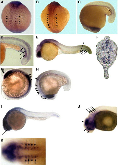
Embryonic expression patterns of zebrafish HS6ST-1 and HS6ST-2 mRNA. (A) 4-somite stage. (B) 10-somite stage. (C) 16-somite stage. Zebrafish HS6ST-2 is expressed in the forming somites (arrowheads in panels A–C). HS6ST-2 expression is down-regulated in more differentiated somites (white arrowheads in C). (D) At 22-somite stage (20 hpf), zebrafish HS6ST-2 is expressed in a stripe of cells in somitic mesoderm (arrows). (E) At 24 hpf, expression of zebrafish HS6ST-2 is detected in the eyes, the brain and ventral medial somites in the tail. (F) A transverse section of the embryo in (E). Zebrafish HS6ST-2 expression is detected in the ventral medial somites and cells surrounding the dorsal aorta (A) and posterior cardinal vein (V) in the tail (as outlined by the dashlines). The level of the section is indicated by the line in (E). (G) At 10-somite stage, zebrafish HS6ST-1 is expressed in the optic vesicles (arrow) and the neural tube (arrowheads). (H) At 16-somite stage, zebrafish HS6ST-1 is expressed in the developing eyes (arrow), the tegmentum, the forming rhombomeres and the neural tube (arrowheads). (I) At 24 hpf, expression of zebrafish HS6ST-1 is detected in the eyes (arrow), the brain and the neural tube. (J) At 33 hpf, expression of zebrafish HS6ST-1 is detected in the diencephalon, the telencephalon, the tegmentum (arrowheads) and in four bilateral patches of cells at the level of the dorsal rhombomeres (arrows). (K) Dorsal view of the embryo in (J), showing the four bilateral stripes of cells in the rhombomeres (arrows). (A–B) Dorsal view, anterior to the top. (C, H) Lateral view, anterior to the bottom. (D–E, I–J) Lateral view, anterior to the left. (G) Lateral view, anterior to the top left. (K) Anterior to the left. |
| Genes: | |
|---|---|
| Fish: | |
| Anatomical Terms: | |
| Stage Range: | 1-4 somites to Prim-15 |
Reprinted from Developmental Biology, 284(2), Chen, E., Stringer, S.E., Rusch, M.A., Selleck, S.B., and Ekker, S.C., A unique role for 6-O sulfation modification in zebrafish vascular development, 364-376, Copyright (2005) with permission from Elsevier. Full text @ Dev. Biol.