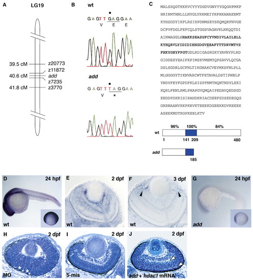
add gene encodes Hdac1. (A) Schematic drawing of LG19. (B) Sequencing of hdac1 DNA isolated from wild type and add mutants. G to T transversion (black squares) replaces 185 E with stop codon in add mutants. (C) Amino acid sequence of Hdac1 and its predicted structure of wild type and add mutant. Nonsense mutation occurs at 185 E (underline) within the Hdac catalytic region (bold letters in sequence, blue in schematic drawing below). Percentage of identical amino acids in zebrafish and human Hdac1 is indicated in each domain. (D-G) Expression of hdac1 mRNA in wild-type (D-F) and add mutant (G) embryos. Arrowheads (F) indicate the expression in the CMZ. Insets (D and G) show embryos at 10 hpf. (H) Retina of embryos injected with hdac1 morpholino oligos. Asterisks indicate folded retinal epithelium. (I) Retina of embryos injected with five mismatch control morpholino oligos. Retinal lamination is normal. (J) add mutant retinas expressing hdac1 mRNA. The formation of the inner and outer plexiform layers are evident (arrowheads).
|
