|
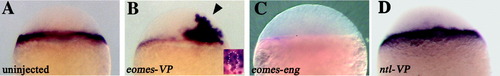
eomes regulates mtx2 expression cell-autonomously. All views are lateral, and all embryos are at sphere stage (4 hours postfertilization [hpf]). Injected construct, if any, is indicated in lower left corner. A: In situ hybridization of mtx2 in an uninjected embryo, showing expression in the marginal cells of the blastoderm and the underlying yolk syncytial layer. B: eomes-VP-injected embryo with ectopic mtx2 expression (arrowhead). Inset shows a portion of the blastoderm of a myc-eomes-injected embryo with Eomes protein expression in the nucleus in brown and mtx2 expression in blue. White outline demarcates a group of cells that coexpress Eomes and ectopic mtx2, indicating a cell-autonomous induction of mtx2 by Eomes. C: Reduced mtx2 expression in an embryo injected with eomes-eng. D: ntl-VP-injected embryo with normal mtx2 expression.
|