
|
Image description by: Tanya Whitfield
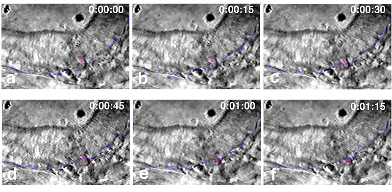
Anatomical structures shown: otic vesicle, rudiment of statoacoustic ganglion
Stage: prim-5 (24h)
Genetic (background) strain: Oxford
Genotype: wild-type
Animal state: live
Labeling: none; DIC optics
Description: Time-lapse video sequence showing a cell delaminating from the floor of the otic vesicle and joining the cluster of cells that form the rudiment of the statoacoustic ganglion. Time specified in h:min:sec. The focus is on the anterior part of the region of delamination, just below the anterior otolith. The coloured drawing over the DIC images identifes the nucleus of the delaminating cell (pink) and the edge of the otic epithelium (blue).
Publication containing this image: Adapted from Haddon and Lewis, 1996 by permission from the Journal of Comparative Neurology.
| Preparation | Image Form | View | Direction |
| whole-mount | still | parasagittal | anterior to right |