Image description by: Tanya Whitfield
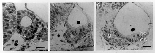
Anatomical structures shown: otic vesicle, statoacoustic ganglion
Stage: 26-somite (22h), prim-10 (28h), long pec (48h)
Genetic (background) strain: Oxford
Genotype: wild-type
Animal state: fixed
Labeling: toluidine blue stain
Description: Origins of the statoacoustic ganglion. A: 26-somite stage–the ventral face of the otocyst epithelium appears irregular, with cells apparently delaminating from the ventral face of the vesicle (arrow), disrupting the continuity of the epithelium and beginning to accumulate beneath it. B: prim-10 stage–formation of the ganglion as a loose aggregation of cells ventral to the ear, beneath the thickened sensory epithelium, which by now shows differentiated hair cells apically and supporting cells basally. C: long pec stage–no longer any signs of delaminating cells; the ganglion (g) has shifted medially and become more compact. hc,sc, hair cells and supporting cells in the anterior macula. Sections taken at the level of the anterior otolith. Scale bar = 25 µm. Reprinted by permission from the Journal of Comparative Neurology.
| Preparation | Image Form | View | Direction |
| LM-section | still | transverse | dorsal to top |

