
- Title
-
Nested expression domains for odorant receptors in zebrafish olfactory epithelium
- Authors
- Weth, F., Nadler, W., and Korsching, S.
- Source
- Full text @ Proc. Natl. Acad. Sci. USA

Spatial organization of the zebrafish nasal epithelium. (A) Scanning electron micrograph of an olfactory rosette of an adult zebrafish. The lamellae depart from a central (nonsensory) raphe. Long, regular, and dense, but sometimes patchy, cilia mark the nonsensory region of the epithelium. Olfactory receptor cells have fewer and shorter cilia and are homogenously distributed on the flat sides in the inner two-thirds of the lamellae (C). (Bar = 100 μm.) (B) Schematic representation of a horizontal cross-section through an olfactory rosette. The lamellae are cut perpendicular to their flat faces. This plane of section is used for all in situ hybridizations. Each lamella consists of two half-lamellae separated by extracellular matrix (gray). The lumen (interface to the water) is surrounded by a black line. The measurement of radial distances (r/ro) is visualized. (C) The dashed line is at relative height 0.60, the cutoff for analysis of radial distances. (Bar = 100 μm.) |
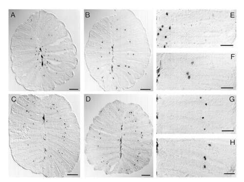
Spatial expression patterns of odorant receptor molecules. Horizontal tissue sections of fresh-frozen nasal epithelium were hybridized with digoxigenylated riboprobes of fZOR6 (A and E), fZOR9 (B and F), fZOR8 (C and G), and fZOR5 (D and H). For orientation, compare Fig. 1. In A–D, the complete sections are depicted (cf. Fig. 1B). (Bar = 100 μm.) (E–H) Photomicrographs taken at higher magnification. (Bar = 50 μm.) Melanophores in the median raphe are visible as elongated dark stain (left edge of micrograph). Labeled cells are small, roundish, and dark. EXPRESSION / LABELING:
|