
- Title
-
Detection of Aeromonas salmonicida subsp. salmonicida infection in zebrafish by labelling bacteria with GFP and a fluorescent probe based on the siderophore amonabactin
- Authors
- Rodríguez-Pedrouzo, A., Cisneros-Sureda, J., Martínez-Matamoros, D., Rey-Varela, D., Balado, M., Rodríguez, J., Lemos, M.L., Folgueira, M., Jiménez, C.
- Source
- Full text @ Microb. Pathog.
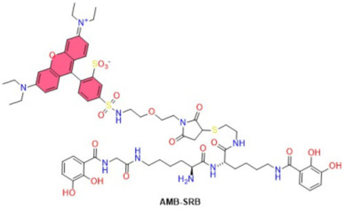
Structure of the fluorescent probe AMB-SRB. |
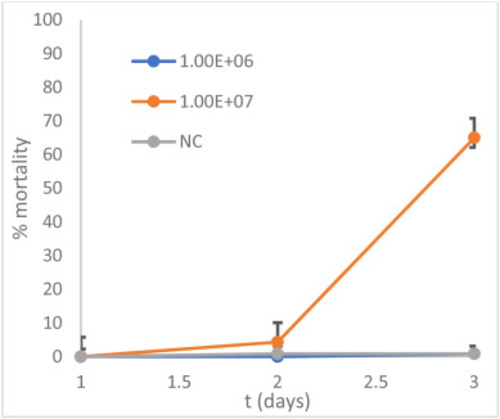
Evolution of the mortality rate of 4 dpf zebrafish embryos exposed to two concentrations of A. salmonicida (106 and 107 bacteria/mL) and the negative control (NC). |
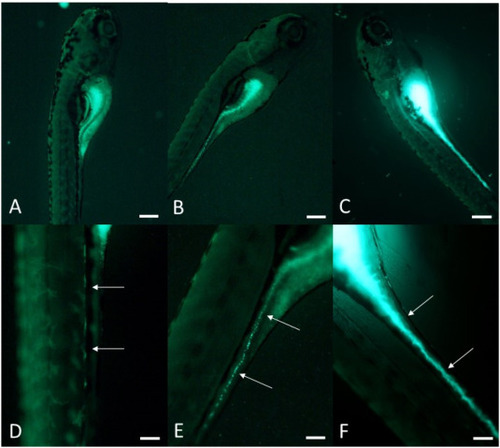
Visualization of zebrafish larvae (Danio rerio) by fluorescence microscopy. A) Control. B) After 24 h of exposure to GFP-labeled A. salmonicida. C) After 48 h of exposure. D) Detail of A at the level of the digestive tract. E) Detail of B at the level of the digestive tract. F) Detail of C at the level of the digestive tract. Lateral views, anterior to the top. Arrows point to the digestive tract. Scale bars: 200 μm (A–C), 100 μm (D–F). |

Visualization of zebrafish larvae (Danio rerio) by confocal microscopy. A–F: Controls. G–L: After 48 h of exposure to AMB-SRB-labeled A. salmonicida. A/D/G/J: bright field. B/E/H/K: Fluorescence. C/F/I/L: Fluorescence and bright field overlay. Lateral views, anterior to the top. Scale bar: 100 μm. |