
- Title
-
Mob4 is required for neurodevelopment in zebrafish
- Authors
- Florindo, C., Mimoso, J.M., Palma, S.L., Gonçalves, C., Silvestre, D., Campinho, M., Tavares, Á.A.
- Source
- Full text @ MicroPubl Biol
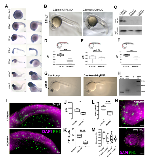
Downregulationof Mob4 in early embryos affects cell proliferation and neurodevelopment: (A) In situ hybridization expression analysis of zebrafish mob4 in wild-type embryos from 90% epiboly, 19, 22, 25, 28, 40, and 48hpf. Bars represent 100 mm. Morpholino-mediated knockdown (KD) of zebrafish mob4 leads to severe neurologic defects during embryogenesis. (B) At 24hpf, morpholino KD of mob4 gives rise to severe neurologic defects. The MHB (arrowhead) is almost absent in 0.5pmol MOB4MO injected embryos, and the eye (e) is smaller. Western blot (C) confirms depletion of Mob4 protein in 24hpf embryos injected with 0.3 and 0.5pmol of MOB4MO but not in 0.5pmol CTRLMO injected siblings. The loading control was carried out using an anti-gamma tubulin antibody (GTU-88). Standard length (D) of 0.5pmol MOB4MO injected embryos is smaller than CTRLMO injected siblings. Trunk length (E) is not affected by 0.5pmol MOB4MO injection. Head length (F) is significantly smaller in 0.5pmol MOB4MO injected embryos. T-test, **** p<0.0001. Bars in A represent 100mm. MOB4 knockout (KO) in CRISPR/Cas9-generated crispants fully recapitulates the MOB4 morpholino phenotype. In no gRNA, Cas9 only, injected F0 embryos (crispants) present a normal phenotype (G) for at 28hpf whereas embryos injected with a gRNA aimed at exon 1 of zebrafish MOB4 gene locus present severe impairment of neural structures. In MOB4 crispants the MHB and the eye (e) are underdeveloped with the phenotype reminiscent of the MOB4MO injected siblings. PCR genotyping (H) confirmed the introduction of an insertion on the zebrafish MOB4 locus in F0 crispants. MOB4 is involved in hindbrain cell proliferation during zebrafish neurodevelopment. (I) Maximum projections of Z-stacks of the hindbrain (yellow line delimited region) of 24hpf embryos where labelling of mitotic cells after PH3 (green) and DAPI (magenta) immunohistochemistry reveals that after injection of 0.5pmol MOB4MO leads to reduced hindbrain area (J) and cell divisions number (K and L, t-test: *p<0.05, ***p<0.003, ****p<0.0002). On the other hand, TUNNEL analysis followed by whole-embryo fluorescent analysis (M) does not indicate significant differences in apoptotic cell number between 0.5pmol CTRLMO and MOB4MO injected embryos. Moreover, co-injection with 0.5pmol p53MO cannot change apoptosis indexes in any experimental group (one-way ANOVA, p>0.05). (N) Maximum projection images of z-stacks of the eye (yellow line delimited) of CTRL and MOB4MO injected embryos labeled with PH3 (green) and DAPI (magenta) show a decrease in mitotic cell number. EXPRESSION / LABELING:
PHENOTYPE:
|