
- Title
-
The Shape of the Jaw-Zebrafish Col11a1a Regulates Meckel's Cartilage Morphogenesis and Mineralization
- Authors
- Reeck, J.C., Oxford, J.T.
- Source
- Full text @ J Dev Biol

Col11a1a knockdown caused a curved body plan and reduced jaw size in zebrafish. Injection of a col11a1a targeting AMO induced a curvature of the body axis, while embryos injected with control AMO developed a straight body axis, (A,B); ((A,B) reprinted under the terms and conditions of the Creative Commons Attribution (CC BY) license (https://www.mdpi.com/2221-3759/8/3/16; Accessed on 15 July 2022) [25]. Normal face and jaw protrusion were present in the control zebrafish (C). In contrast, the morphant zebrafish demonstrated a reduction in face and jaw protrusion. Heart edema was also observed, (D). Alcian blue staining of the pharyngeal arch cartilages showed normal patterning and distally projecting cartilages (E), while Alcian blue staining in the morphant showed perturbed pharyngeal cartilage patterning characterized by a decreased protrusion of the 1st arch and a posteriorly facing 2nd arch, (F). Scale bar in (A,B) 0.5 mm; Scale bars in (C–F) 50 μm. (m; Meckel’s cartilage; pq; palatoquadrate; ch; ceratohyal; cb; ceratobranchial). PHENOTYPE:
|
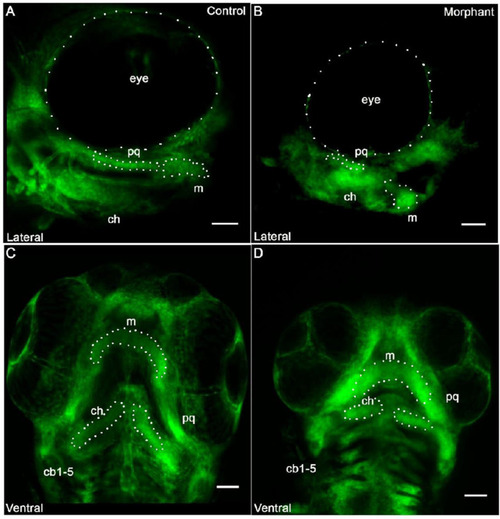
Cranial neural crest derived cells form segmented pharyngeal arches with abnormal shape under knockdown conditions of col11a1a. Fli1a:EGFP positive cells indicated that the neural crest derived cells were present within the pharyngeal arches. After occupying the pharyngeal arch, however, cells failed to form proper morphology. While the cells aligned to form an elongated structure in the palatoquadrate (pq) that joins the Meckel’s cartilage in the control morphant (A), the pq of the col11a1a morphant did not form the extended structure and the Meckel’s cartilage did not extend distally (B). The ventral view indicated segmentation of each of the developing pharyngeal arches in both the control (C) and the morphant (D). However, altered morphology was observed in the morphant (D). Scale bar 50 μm. (m; Meckel’s cartilage; pq; palatoquadrate; ch; ceratohyal; cb; ceratobranchial). PHENOTYPE:
|
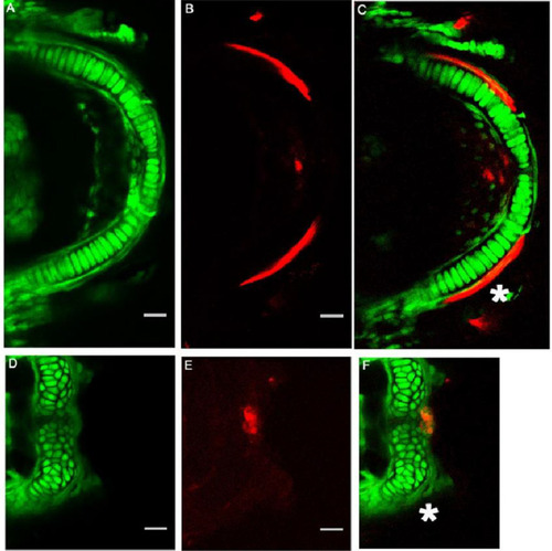
Col11a1a expression was required for Meckel’s chondrocyte organization, extension, and mineralization. The Fli1a:EGFP expressing cells in the Meckel’s cartilage formed an organized stack of chondrocytes that allowed the arch-like structure of Meckel’s cartilage to form (A) Alizarin Red staining indicated that the perichondraal cells produced a mineralized structure immediately adjacent to the Meckel’s cartilage ((C), white asterisk) and initiated mineralization at the central distal tip (B,C). In contrast, the cells of the morphant were not flattened, nor were they organized into a stack of cells as observed in the control ((D) compared to (A)). Lateral mineralization of the bilateral rods in the morphant was not detected ((F), white asterisk). Mineralization was detected by Alizarin Red staining at the central distal tip (F). Scale bars 20 μm. PHENOTYPE:
|

Col11a1a knockdown disrupts the organization of bone forming cells that line the Meckel’s cartilage. Ventral view of sp7:EGFP positive cells in 5 dpf control zebrafish (A,C) and morphant (B,D). sp7:EGFP expressing cells line the cartilage template in the control zebrafish (C). sp7:EGFP expressing cells form small condensations at the mediolateral cartilage but fail to occupy the location of future mineralization immediately adjacent to the Meckel’s cartilage as observed in controls (D); Scale bar 20 μm. PHENOTYPE:
|
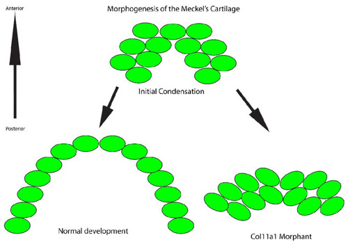
Model of Meckel’s cartilage development and the cellular organization in normal development and in the col11a1a morphant. During normal development, the cells (green) reorganize and form a single file stack of cells that promotes extension distally. In contrast, inhibition of col11a1a expression prevents reorganization into stacked cells. Therefore, the Meckel’s cartilage does not extend distally. |

Model of Meckel’s cartilage mineralization in the col11a1a morphant. The organized chondrocytes (green) serve as a template for bone forming cells (red) to generate a calcified matrix. These cells extend along the template and form two bilateral mineralized rods. Therefore, perichondral cells cluster and produce abnormal mineralization at the cartilage template. |